
Ever noticed how one picture can tell you so much about your pet’s health? Vets use cool tools like digital X-rays and ultrasounds (special scans) to see inside your pet’s body. It’s like getting a sneak peek at what’s happening under the fur.
These images help spot little issues early, like tiny cracks or changes that might mean trouble. And that means we can plan care to keep your furry friend happy and healthy. Stay with us to learn how these tools make pet care even better.
Understanding Diagnostic Imaging Techniques in Veterinary Medicine
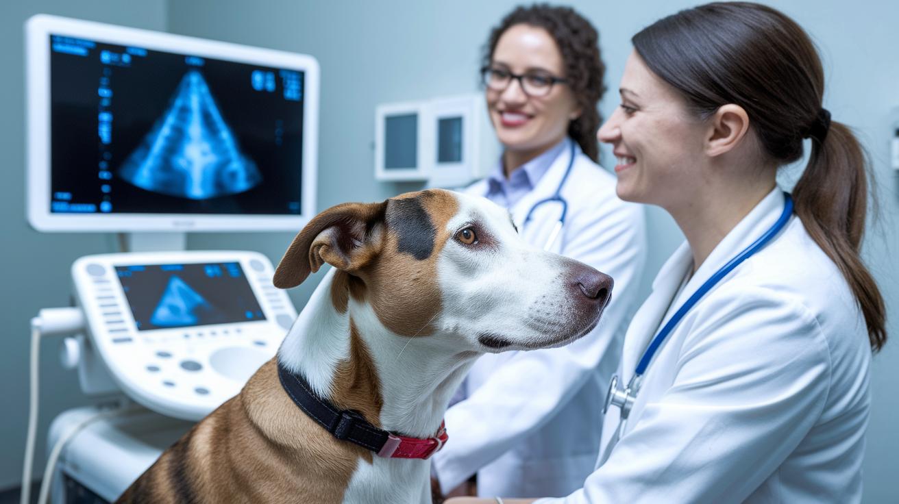
Veterinary imaging is all about using cool, high-tech tools to peek inside your pet safely. Digital X-rays (like a careful snapshot of bones and tissues) give us clear images that help spot fractures or signs of sickness in areas like the chest, tummy, or even the teeth. It’s kind of like taking a neat photo that reveals hidden details inside your furry friend.
Ultrasound works a bit like a live movie. It uses sound waves to create moving images, so you can see a tiny heart beating or a kitten in the early stages of life. This tool is great for checking soft tissues in the belly and catching any problems early.
CT scans build up layers of pictures to form a 3D view of your pet. This method is especially handy when looking at tricky spots like a pet’s brain or complex bone structures. By showing different angles, CT scans give vets a full picture of what might be wrong.
MRI uses strong magnets to make very detailed images of soft tissues such as muscles and the central nervous system (the body's control center, like the brain and spine). This helps with examining delicate parts and catching any small changes before they become bigger problems.
Together, these imaging tools work as a complete set, allowing vets to check for broken bones, examine soft tissues, and monitor nerve health. This way, every pet gets the careful attention they need, with images that truly capture the details of their well-being.
Radiographic Techniques and X-Ray Procedures for Animal Diagnostics
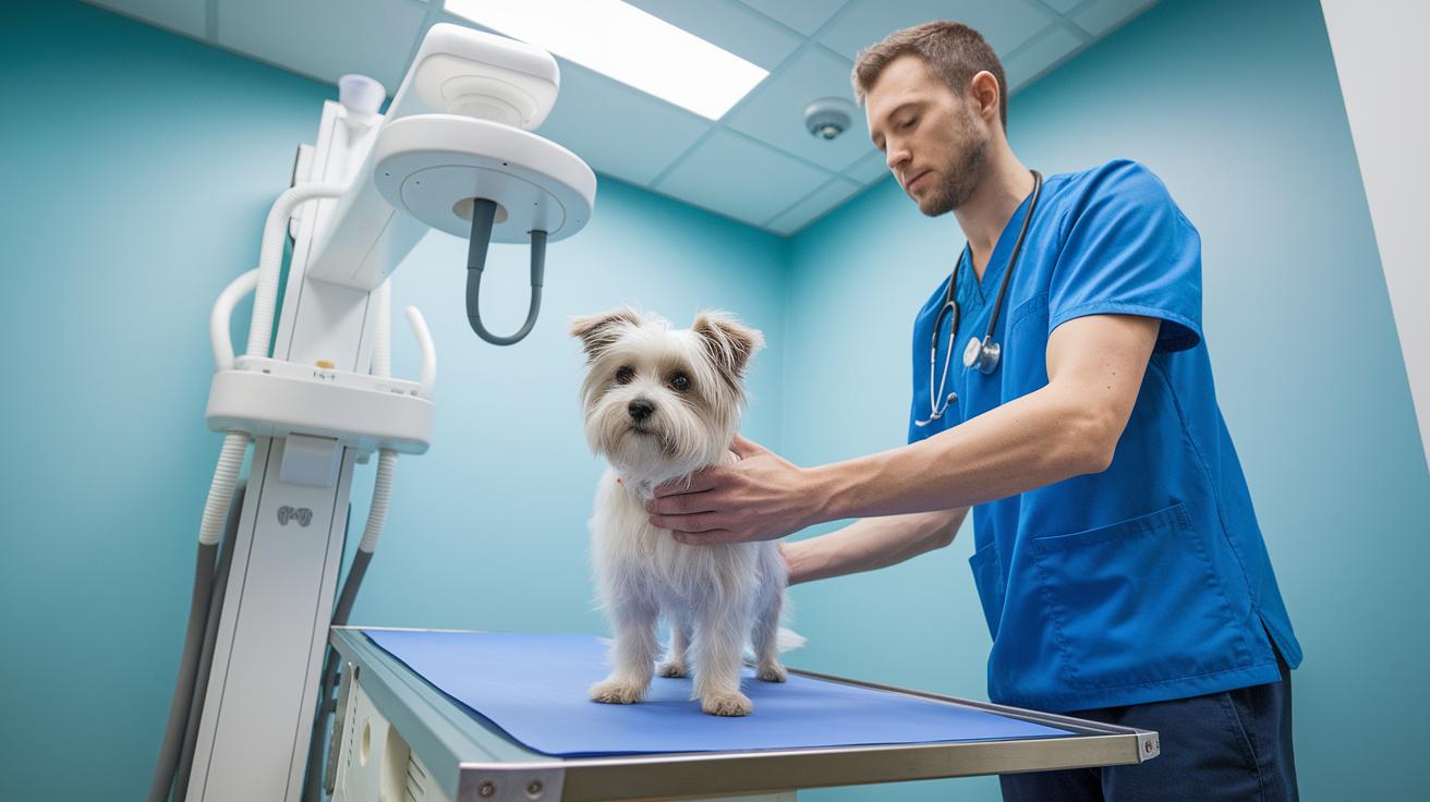
Radiography is like using a special light to create flat pictures of your pet's bones and soft tissues. When X-rays pass through your pet’s body, different parts soak up the energy in their own way. This process makes a shadowy image that shows the differences between structures. And did you know? A high-resolution detector can catch details as tiny as a hairline fracture, revealing clues to an animal's injury that might otherwise go unnoticed.
Today’s X-ray machines use smart digital software (computer programs that make images clear and process them fast) so veterinarians can easily spot fine details in your pet's chest, tummy, teeth, and bones. They also include strict safety steps like monitoring the radiation dose (the amount of energy used) and reducing extra exposure. So, whether it’s a quick checkup or a closer look at a possible bone issue, every exam is set up to protect your beloved pet.
All these tools and safety measures work hand-in-hand to make sure X-ray exams are both effective and safe, giving clear and reliable images that help vets decide the best care for your furry friend.
Companion Ultrasound and Doppler Imaging in Veterinary Practice
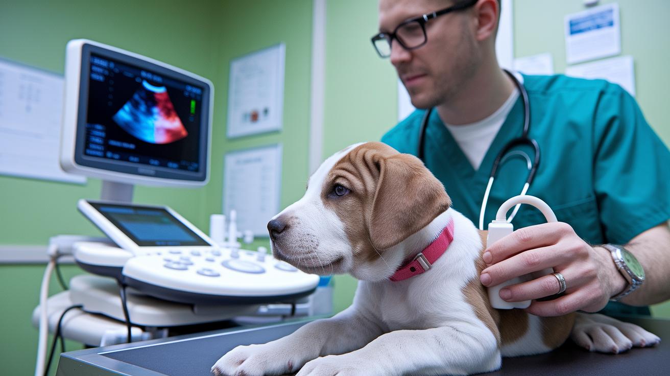
Ultrasound uses high-frequency sound waves to create live images of soft tissues. Imagine watching your pet's heart gently beat or spotting a playful kitten’s belly move. It’s like having a tiny TV inside your pet, letting vets see organs like the liver, kidneys, or heart very clearly.
Color Doppler adds a fun twist by checking how blood flows through your pet’s vessels. Think of it as a colorful traffic report, showing where blood is rushing and where it might be slowing down. This technique helps vets find vascular issues whether it’s a small pet like a cat or dog, or a larger animal like a horse.
Portable clinical units bring this cool technology right to your pet. A mobile ultrasound cart can be rolled straight into a clinic, or even taken out into the field, to check abdominal, heart, and reproductive health without any delay. For instance, one vet used a portable unit to capture a puppy’s heart in real time, catching a mild abnormal rhythm before it became a bigger concern.
This easy-to-use, advanced imaging tool makes routine checkups and emergency scans both faster and more accurate, giving every pet a better chance at a healthy, happy life.
Computed Tomography Applications: Fast 3D Imaging and CT Processing
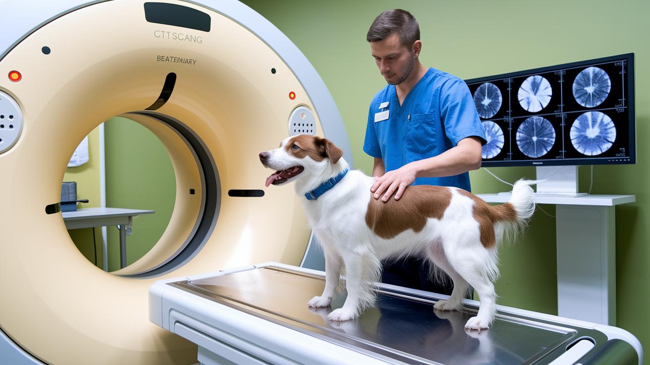
Computed tomography (CT) uses X-rays and simple computer math to make cross-sectional pictures of your pet’s body. The scanner moves around your pet, taking lots of tiny images. Then the computer pieces them together, much like a photo puzzle, to show you a detailed 3D view of both bones and soft tissues. It works well whether it’s a small dog or an animal as big as a horse.
When scanning smaller pets, the CT machine adjusts its settings, lowering the X-ray strength and making thinner slices, to match their size. For bigger animals like horses, a wider scanner is used to cover every part of their larger body. Imagine snapping many little pictures and then joining them to make one big image; that’s exactly how it works.
The CT scan process has a few clear steps. First, image segmentation separates different body tissues so each can be studied on its own. Next, the computer builds a 3D model by combining these segments together. Then, volume rendering brings out all the details, much like stacking thin layers of cake to reveal the full flavor. One technician put it simply: “Each slice is like a thin cake layer – stack them just right, and you get the whole picture!”
These steps help vets check on heart, brain, and bone conditions quickly and accurately. With fast scans and speedy processing, CT scans are a powerful tool in keeping your pet healthy.
Magnetic Resonance Imaging for Small Animals and Equines
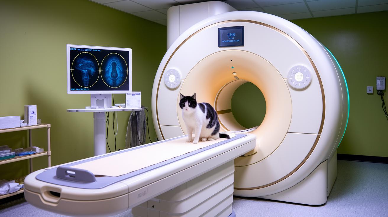
Small Animal 1.5T MRI machines are built especially for our furry friends. They don’t use helium and are designed to run almost flawlessly during scans. They check the strength of magnetic fields to produce clear and detailed images, and clever noise suppression cuts out unwanted background signals so you can see soft tissues with sharp contrast.
These systems come with a feature that works like a built-in camera stabilizer. Even if a pet moves a bit, the smart technology adjusts in real time so every image stays crisp. This is great when scanning a restless cat, as it helps prevent any blurriness.
Advanced software steps in to fine-tune the images by aligning and processing the data with care. This makes it easier for technicians to spot tiny differences in soft tissue and nerve structure. By cutting down on extra artifacts (stray signals), these scans give reliable results for checking both the brain and other soft tissues in small animals and horses.
New hardware options also bring a variety of coil setups that improve signal capture. This extra detail helps vets make earlier diagnoses and plan treatments more accurately. In short, the blend of solid engineering and innovative software delivers high-resolution images that pet owners can count on for accurate care.
Every scan gives vets a clearer view of your pet’s organs, guiding them to choose the best interventions to keep your loved ones healthy and happy.
Case Studies in Veterinary Imaging Diagnostics
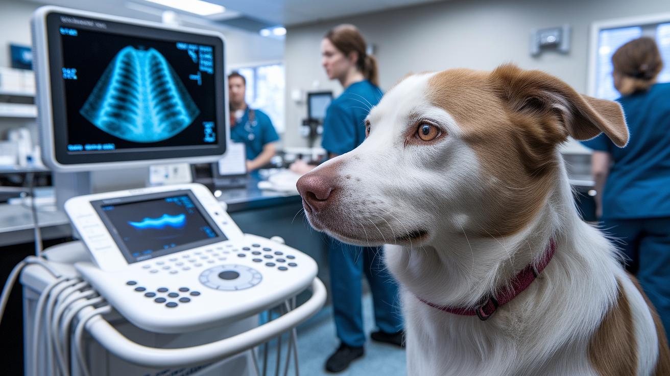
Imagine a bustling day at the clinic where small clues help decide the best imaging test. Take the playful Labrador Retriever with an upset stomach, an abdominal ultrasound was done that showed important details about his insides. Then, a wise old Staffordshire Terrier with a nagging cough had a chest x-ray, and the pictures helped the team spot hidden lung issues.
Over in the horse area, a 2-year-old Thoroughbred with a slight limp had a standing CT scan. The detailed 3D images pinpointed a minor joint problem so that treatment could start quickly. Next, a 3-year-old Domestic Longhair acting a bit strangely and with signs of seizures received a brain MRI. Its clear pictures helped the vet decide on the next steps.
For dental care, a 13-year-old Domestic Shorthair had dental x-rays to check for tooth resorption. In another case, a 10-year-old Shih Tzu showing signs of chest discomfort got a thoracic CT scan that revealed a mass in his lung area, leading to further specialist review. Lastly, a 2-year-old Dachshund suffering from severe back pain underwent a spinal MRI, which identified an intervertebral disc problem and guided targeted treatment.
| Animal | Age | Imaging Modality | Key Finding |
|---|---|---|---|
| Labrador Retriever | 1 year | Abdominal ultrasound | Chronic vomiting |
| Staffordshire Terrier | 13 years | Chest radiograph | Persistent cough |
| Thoroughbred | 2 years | Standing equine CT | Lameness |
| Domestic Longhair | 3 years | Brain MRI | Seizure workup |
| Domestic Shorthair | 13 years | Dental radiography | Tooth resorption |
| Shih Tzu | 10 years | Thoracic CT | Mass detection |
| Dachshund | 2 years | Spinal MRI | Disc extrusion |
These real-life examples show how each imaging test helps guide accurate and thoughtful care for our furry friends. The tests provide strong evidence so vets can quickly decide on the best treatment to help pets feel better.
Emerging Trends and AI Integration in Veterinary Imaging
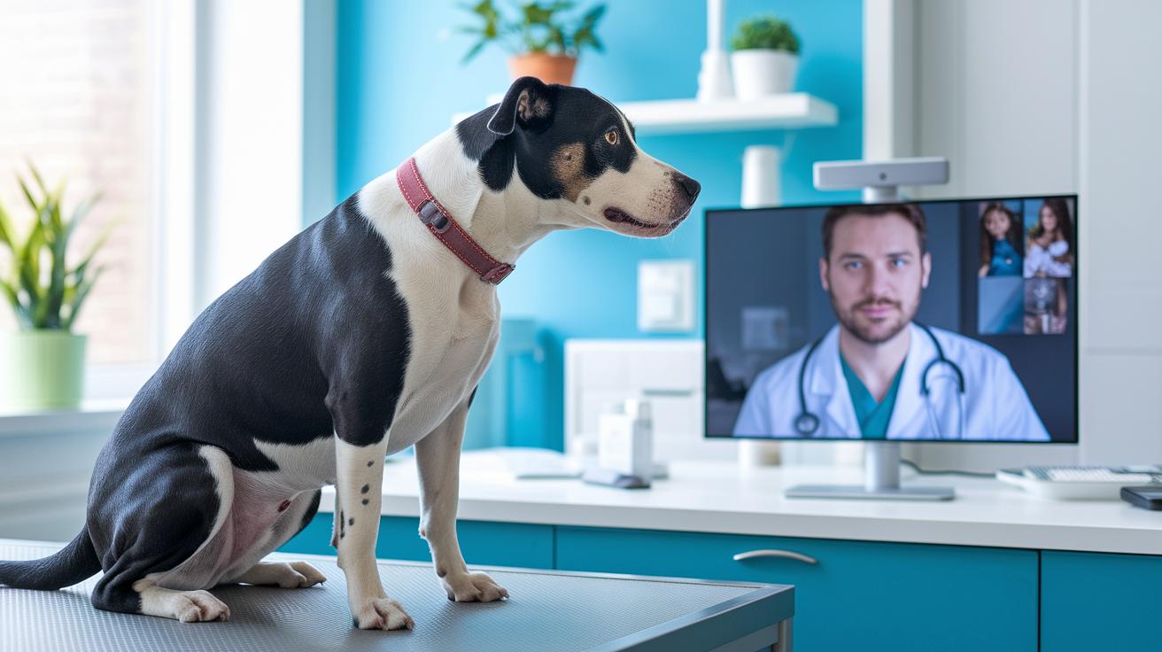
AI is giving pet imaging a whole new look. Smart computer programs adjust images in real time, making them clearer and speeding up scans. With systems that let vets check images remotely via virtual consultations, help is always just a click away, even if the expert isn’t in the same room. These tools sift through data quickly, letting vets pinpoint issues fast, so both your pet and you benefit from quicker, more precise care.
• Brighter, sharper images
• Shorter scan and anesthesia times
• Automatic spotting of unusual spots or lesions
• Easy remote access and virtual chats with experts
• Organized reporting and secure cloud storage
All in all, these AI improvements are transforming pet care into something smarter and more accessible, helping every pet get the best possible care.
Final Words
In the action of veterinary imaging, we explored how radiography, ultrasound, CT, and MRI work to give clear insights into pet health. We shared real pet examples and covered AI-driven advances that make a real difference in care. Small animal and equine cases showed how these methods help with accurate diagnoses and smoother recovery. It’s a bright time for pet care, sparking new confidence in making smart decisions for each special companion.
FAQ
What is veterinary imaging near me?
The term veterinary imaging near me refers to local diagnostic services like X-rays, ultrasounds, CT scans, and MRIs that your nearby veterinary clinic offers to help detect and manage your pet’s health issues.
What is a veterinary radiology website?
The veterinary radiology website provides trusted online resources where pet owners and professionals can learn about imaging services, access case studies, and find contacts for advanced diagnostic care focused on animal health.
What does veterinary radiology interpretation involve?
The process of veterinary radiology interpretation involves reading and analyzing diagnostic images to help veterinarians accurately diagnose conditions, making it easier to plan treatments and improve your pet’s overall wellbeing.
What is Hallmarq veterinary imaging known for?
The name Hallmarq veterinary imaging is associated with high-quality imaging solutions; it represents a brand that uses advanced technology to produce clear images, aiding in the accurate diagnosis of pet health concerns.
What is the Veterinary Imaging Association?
The Veterinary Imaging Association is an organization dedicated to advancing pet care by promoting quality imaging standards, sharing educational resources, and supporting professionals in the field of veterinary diagnostic imaging.
What is diagnostic imaging in veterinary medicine?
Diagnostic imaging in veterinary medicine uses techniques like radiography, ultrasound, CT, and MRI to visualize internal structures, ensuring that veterinarians detect issues early and create effective treatment plans for pets.
What role does a veterinary radiology technician play?
The veterinary radiology technician operates imaging equipment, ensures quality picture capture, and supports the radiologist’s analysis so that accurate, safe, and reliable diagnostic images are produced for proper pet care.
How does health veterinary imaging benefit pets?
Health veterinary imaging benefits pets by providing clear internal images that help detect fractures, soft-tissue problems, and other issues early, allowing for quicker diagnoses and more effective treatment planning for your pet’s comfort.